
Plachta lab uses live-embryo imaging to elucidate important role of actin filaments early embryo mitosis
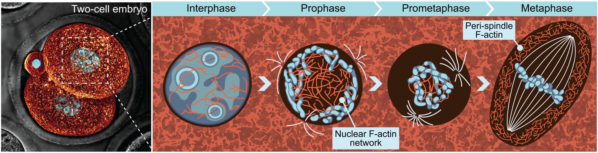
A new paper from the Plachta lab used live-embryo imaging to discover a contractile network of nuclear actin filaments that organize chromosomes during the early stages of mitosis. Additionally, they observed a network of actin filaments enclosing the metaphase spindle which prevents uncontrolled spindle elongation. Their work provides an explanation as to how the early mouse embryo is able to achieve faithful chromosome segregation in the absence of canonical mitotic machinery.
You can read their full paper, as published in Science, here.